|
| |||||||||||||||||||
The Gold Standard of primary hip replacement, 36 years on
Clinique de l’Yvette - 91160 Longjumeau - France
The Low Friction Arthroplasty designed by John Charnley (1911-82) goes back to 1962.
In November 1962, after in-depth research into cemented implant fixation
and into bearing surfaces, and following the unhappy experience with Teflon
cups (1959-62), John Charnley implanted his first low friction arthroplasty
(LFA). For the first time, he was using a cemented high density polyethylene
(HDP) cup, which articulated with a stainless steel femoral head of 22.25 mm
(7/8") diameter. The principle of the LFA has remained the same ever since
(Fig. 1).
The first article, by the LFA’s designer, was published in the
Journal of Bone and Joint Surgery (British version), in 1972.1 It gave the
radiographic and clinical results of 379 LFAs, out of a total first series of
582 primary hip replacements performed at Wrightington between November 1962
and December 1965 (an annual rate of nearly 200 hip replacements, even in
those early days). In other words, 65% of the patients who had received an LFA
had been followed up, for between 4 and 7 years only - a coverage that would
not really be acceptable nowadays. While all the stems used in that initial
series were of the conventional monobloc flat-back design, made of steel, and
fixed with cement, the pattern on the acetabular side was less homogeneous:
159 cups (29%) were cementless metal-back press-fit devices, which could
rotate in the acetabulum around an HDP spigot. These cups did less well in the
short term, as compared with the cemented cups that had been implanted over
the same period of time, and were therefore no longer used after the end of
1965. Charnley’s disciples2 maintain that it was this set-back that finally
decided Charnley to use cement with all acetabular components.
Thirty-six
years on, the LFA remains the gold standard, world-wide, the benchmark against
which all other hip replacements are judged. It is unrivalled in its
universality and longevity. It was an immediate success, and came to be used
on a very large scale: it is thought that, to date, a million LFAs have been
performed world-wide. However, the design was copied many times, and many
combinations were devised (Charnley-Müller, Charnley-Kerboull, etc.), to the
point where the term LFA has become a little imprecise.
We have tried to
review the literature of papers and abstracts published over the past 5 years
(see References), in order to establish a somewhat clearer picture. The
present article is deliberately confined to the results of large, general
series; studies of specific patient populations (RA; patients under 50 years
of age) have not been included, since they would merit an article of their
own. It is hoped that this meta-analysis of the outcome of the LFA (in its
original form and its further developments) in the 1993-98 time-frame will be
accepted in the spirit in which it is proffered - as a modest and objective
contribution to our state of knowledge that respects the different authors and
their opinions.
OF THE ORIGINAL
CHARNLEY LFA
The 1995 International Symposium at Lyons
This anniversary symposium to celebrate "Charnley Total Hip Arthroplasty: 33 Years of World-Wide Experience" was organized by A.C.O.R.A., the Rhone-Alpes Association of Orthopaedic Surgeons, under the chairmanship of C. Picault. It provided an opportunity for international stock-taking, to see how the LFA had fared. No fewer than 15 countries were represented: ten of the participating countries were European; however, North America (US, Canada), Japan, Australia, and Mexico also took part.
Points of agreement
The volume of abstracts3 shows that there was strong agreement on certain
points, which are summarized below:
- The clinical results of the "original" LFA, after 20-25 years, are universally considered as satisfactory by the users;
- The stem should be made of high-strength steel;
- The overall incidence of aseptic loosening is higher on the acetabular than on the femoral side (except for the results presented by Munuera & Garcia-Cimbrelo, La Paz Hospital, Madrid, Spain);
- Aseptic loosening is often clinically silent for a long time, or well tolerated;
- Risk factors for loosening are young patient age, and - to a lesser degree - male gender, and a high activity level;
- Polyethylene (PE) wear is the key factor that governs the service life of the implant;
- In order to respond to the universal wish to delay the production of wear debris and the resultant osteolysis, better-quality PE will have to be developed;
- The final conclusion was drawn by M. Porter (UK), who, in the light of an estimated annual failure rate of 1/1000, advised his colleagues to use the cemented LFA with an Ortron stem and a dorsal flange (Elite), and the flanged Ogee socket.
Points of disagreement
- Surgical approach: The original technique involving a trochanteric osteotomy, much favoured by Charnley (Wroblewski, Clarac, Kerboull) is no longer in universal use: some surgeons have adopted an anterolateral approach (Picault, Hardinge), or a posterolateral one (Michel, Durandeau et al, Nixon et al).
- Canal fill: some surgeons preserve the cancellous bone (Draenert et al, Picault, Brady et al), while others will sacrifice this bone and use bulkier implants, with a thinner layer of cement (Kerboull).
- Cementing technique: "old-fashioned" (Picault), using cement of standard viscosity (Hernigou et al, Hodgkinson); 2nd-generation (Johnston, Callaghan et al, Kawai et al, Carlsson et al); and even with vacuum application (Draenert).
- Stem modularity: advocated by some (Wroblewski, Le Saout et al), but considered dangerous by others (Johnston and Callaghan, Clarac, Salvati, De Nayer et al).
- Femoral head material: alumina ceramics (Wroblewski) and zirconia ceramics (Cales, Le Saout et al) are now coming in.
- Cup fixation: "classical" cement fixation (Clarac, Kerboull); cemented with a flange (Hodgkinson, Wroblewski, Porter); cemented with spacers (Caton); and even cementless (Prudhon, Callaghan).
As will be seen, there are still many question marks, and the solutions proposed vary widely.
The overwhelming majority of the clinical series presented at Lyons had been published previously in the international literature. In this article, little reference will be made to these earlier publications. However, the question arises of what has been happening over the 3 years since the Lyons symposium. In order to obtain a more up-to-date picture, it was decided to review the literature since 1995, to gather more recent results by authors who had attended the symposium, as well as results from teams that had not attended the Lyons event.
Results published since the Symposium
Surgeons from the Cochin Hospital were among the speakers at Lyons; since then, they have presented, to the 1995 SOFCOT meeting, a study of original LFAs performed by one surgeon in the time between 1970 and 1973.4 This study showed an overall 20-year implant survival rate of 93%, and concluded that the functional results were good; however, the long-term outcome was threatened by osteolysis as a result of PE wear debris and foreign body granuloma formation. The A.C.O.R.A. group, who had organized the Lyons event, saw the publication, in 1996, of a Lyons University thesis by MS Marduel-Picault, which summed up the experience, over more than 20 years, of the Group’s founder members. The general conclusion was that everything was just fine; hard statistics were, unfortunately, less in evidence.5
Results published by Lyons "absentees"
Seven publications giving the long-term results of the original Charnley
LFA have been selected for purposes of this article. Salvati represented the
New York Hospital for Special Surgery (HSS) at Lyons, but did not present
their results. The HSS results obtained with the original LFA had been given 3
years earlier, at the 1992 SOFCOT meeting, by Msika.6 In the light of 82%
survival after 17 years, the authors recommended cemented THR for patients
aged over 65 years, with a life expectancy of around 15 years. Karachalios et
al (KAT Hospital, Athens) had presented their 12-18-year results of 95
original LFAs implanted from 1973 to 1979, in a paper published in 1993.7 They
insisted upon the importance of anatomical placement of the cup in the
interest of long-term implant survival. One of the most notable absentees from
the Lyons symposium was the Mayo Clinic, of Rochester, Minnesota. The
follow-up studies by that centre of the first 333 LFAs have been amply
documented by Coventry, Kavanagh and others. The latest up-date, of 1994,8
showed an overall probability of revision of 11% at 15 years, which rose to
16% at 20 years (closely matching the results of the New York HSS). The
authors also stressed the influence of age at the time of the initial THA: the
risk of revision was 12% for those over 70 years of age, versus 27% for those
less than 59 years of age. Lars Neumann’s group (Odense, Denmark) was also
among the absentees at Lyons. In 1994, this group had reported a 10.7%
probability of revision at 20 years, in a follow-up of 241 LFAs performed by
one member of the team (KH Sørensen) between 1968 and 1974.9 The Swedish
results obtained by Herberts and Malchau (Garellick et al10) were obtained in
a series of 95 original LFAs implanted between 1973 and 1977: implant
survivorship was 92% at 10 years, but down to 83% at 16 years (matching the
pattern observed at the HSS). In France, P. Gardes, in a follow-up study of
100 LFAs (out of 223 THAs performed, by one surgeon, between 1974 and 1976)
stressed the occurrence of PE wear in the younger patients.11 The Philadelphia
group (Rothmann and Hozack), at the 6th Annual Meeting of the AAHKS in 1996,
presented their follow-up of 1157 original LFAs. They reported an overall
probability of non-revision at 20 years of 81% (stem: 87%; cup: 87%), but
probabilities of definite loosening of 23% and 30%, respectively, for the stem
and the cup.12 In 1997, Kobayashi et al13 published their results of 296
primary LFAs implanted, at Shinshu University, between 1972 and 1984. At 16
years, the femoral component survival rate was 95.6% using revision as the end
point, and 91% using radiographic evidence of failure as the end point.
Survival was found to be adversely affected by large width of the femoral
canal. A report from the Swedish Hip Arthroplasty Register14 of 11,880
Charnley LFAs (original pattern and Evolution) implanted in OA patients in
Sweden between 1979 and 1986 showed a 14% overall probability of revision for
aseptic loosening after 17 years.
While these studies do not fundamentally
alter the conclusions of the Lyons symposium, they show a change, in the sense
of a doubling of the rate of poor results around the 20-year mark. Results are
clearly influenced by the number and the level of experience of the surgeons
operating on the patients in a given study: where hip replacements were
performed by senior surgeons or a single surgeon, results are consistently
better than in studies involving several surgeons of greater or lesser
experience. The final conclusion concerning the original LFA is that the
device has a "gold standard" overall revision rate of between 10% and 20% at
15-20 years’ follow-up; and that young patient age and PE wear will affect the
results adversely, in the long term.
OF THE ORIGINAL
CHARNLEY STEM
Long-term results of the original flat-back stem
The results described above were obtained with the original flat-back stem designed by John Charnley. This monobloc stainless steel stem was comparatively flat in the coronal plane, with fairly sharp edges. Since the Lyons symposium, two of the participants have reported their results with this stem pattern. The experience of Poitiers University Hospital was described by Soyer et al15, in 1997. In a series of 309 primary hip replacements performed, between 1972 and 1975, by one surgeon (JP Clarac), using Charnley’s original technique (trochanteric osteotomy; first-generation cementing), the probability of stem survival at 20 years was found to 66% using revision for whatever reason as an end point, and 84% using femoral component loosening (migration, complete radiolucent line, or stem fracture) as the end point. Risk factors were implantation in varus, male gender, and a reasonably high level of activity. Garcia-Cimbrelo16 followed up 680 LFAs performed between 1971 and 1979, and found a probability of femoral osteolysis developing after 20 years of 11%. The author also found a stem fracture rate of 4.3%. However, in his study, neither age nor activity were found to be statistically significant risk factors; significance was shown only for implantation in varus. Unlike other authors, Garcia-Cimbrelo found the cup to do better than the stem in terms of overall outcome (11.5% probability of revision, as against 14%).
From the original stem to the Elite
Starting in 1969, all the Charnley stems made by Thackray (Leeds, UK) have
had a matt finish applied by a Vaquasheen process, rather than a smooth
surface. However, the chief reason that prompted John Charnley to change the
shape of his original flat-back design was concern over stem fractures. While
in 1975 he found only 17 fractures in a total of 6,500 stems implanted
(0.26%), the rate had gone up with time (1962-72: 2 fractures; 1972-74: 8
fractures). This finding led to the creation, in 1974, of the Evolution range.
The fracture rates reported in the literature were between 0.32% and 19%,
which is why Charnley’s followers were so frequently faced with the problem
that they published a paper, in 1994,17 on 125 cemented revisions for
fractured stems (of which 118 Charnley flat-backs); stem survivorship at the
end of 11 years was found to be 90%.
In order to improve the fatigue
strength of the stem, Charnley had essentially wanted to increase the volume
and the cross-sectional area of the device. This led, in 1974, to the
round-back stem, which was very much stiffer than the flat-back. However,
Charnley was well aware that stem fracture was related to loosening; he
therefore decided to improve the quality of stem cementing, by the
introduction, in 1975, of anteroposterior flanges to prevent the escape of
cement at the neck resection level and to pressurize the cement in the femoral
canal. Stainless steel was replaced by Ortron 90, in 1982, and the neck
circumference was reduced to 10 mm. This resulted in the creation, in 1984, of
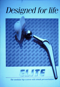
the Charnley Evolution range, and, eventually, in the modular Elite stem
introduced in 1986 (Fig. 2). By then, Charnley had been dead for four years,
and it was his disciples, first and foremost Mike Wroblewski, who were
responsible for the latest innovations.18
What, then, were the effects of
stiffening the stem? We have found only one study, performed jointly by
American and South African authors, that deals specifically with this
question.19 In this study, the fate of 264 LFAs with first-generation stems
(1970-71) was compared with that of 402 LFAs with second- to fourth-generation
stems (1975-86). The results were somewhat heterogeneous. In terms of fracture
risk, there was a marked improvement, with a probability at 10 years of
survival without stem fracture of 94.6% for the flat-back stems, and of 99.6%
for the stems with a larger cross-sectional area. However, the probability at
10 years of revision because of aseptic femoral component loosening was 0.7%
for the original stem, and 13.2% for the bulkier stems. This stands to reason
if one considers that flat-back stems fracture subsequent to loosening;
however, the rate still appears abnormally high, especially considering that
the Evolution stems had been inserted using second-generation cementing
techniques. It is unlikely that the textured (matt) surface finish of the
stems with the larger cross-sectional area (as opposed to the polished finish
of the earlier stems) was the sole factor responsible for the marked increase
in the rate of loosening observed.
 |
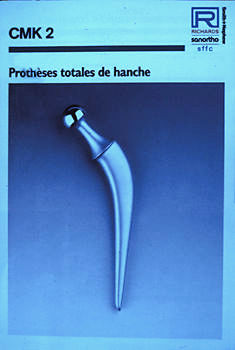 |
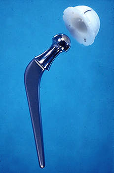
| Figure 1 : LFA classic | Figure 2 : CMK 2 |
BY OTHER DESIGNERS
Wrightington designs
After Sir John’s death, and while Wroblewski was developing the Evolution device faithfully following his master’s stem concept, a plot of truly Shakespearean proportions was being hatched right there at Wrightington. Kevin Hardinge, who had already abandoned the sacrosanct trochanteric osteotomy, was developing the Wrightington Frusto-Conical (WFC) hip replacement, which came out in 1981. The battle lines were drawn up, and 260 monobloc LFA Evolution (Ortron, flanged) stems pitted against 260 WFC stems with a larger, circular cross-section with longitudinal flutes and shaped like a segment of a cone. The results were presented by Hardinge at Beaune (EHS ‘98), and published by Sochart and Hardinge in July 1998.20 Ten-year survivorship, with revision for aseptic loosening as the end point, was 99% for both stems. However, at 15 years, the Charnley was seen to have been stabbed in the back by the young pretender: stem survivorship was only 87% for the Charnley design, as against 98% for the Wrightington FC. These results may, however, have been affected by other factors, such as the surgical approach and cementing techniques employed. The Charnley had been inserted following trochanteric osteotomy and without a cement restrictor; for the WFC, a Hardinge approach and advanced cementing had been used. These technical differences should be considered in weighing the results of the survival studies.
French designs
While Charnley look-alikes and clones of the original LFA and the Evolution family were mushrooming world-wide, the French market came up with several stems derived from the LFA concept.
The Charnley-Kerboull stem
This stem first appeared in 1972, in the monobloc CMKI and later CMKII versions (Fig. 3). This device had an angle of 130° (instead of that of 125° of the Charnley stems); above all, the stems were thicker all along their length, in order to produce greater canal fill with a stem " pressed" into the femur. As a result, there was a thinner cement mantle; in fact, in some zones, the mantle was almost non-existent. Also, many different models were designed, so as to adapt the implant to the different anatomical patterns encountered. The design rationale of the Charnley-Kerboull stems is discussed in detail in a book21 published by the Cochin Hospital in 1985. Flanges were omitted, as a matter of policy, both from the stems and from the cups. At the 1994 SOFCOT meeting, Dejean22 presented the results, at a mean follow-up of 15.5 years, of 70 THRs performed by Kerboull himself, between 1975 and 1977. Two stems (2.8%) had fractured, but only one was revised; another 2 stems were considered to be loose, but were not thought to be causing sufficient clinical problems, at the time of reporting, to warrant revision. Thus, the stem loosening rate observed was 5.7% at 15-16 years. The monobloc polished 306 LVM stainless steel CMKIII was introduced in 1987.
The French modular stems
In 1988, Benoist Girard brought out the Kerboull CMKIII, with a stem and
femoral head in titanium. This metal had been chosen in order to allow
cemented Charnley-style stems and femoral heads to be manufactured; however,
as shown by Langlais23 in 1993, this was not an entirely safe option. Titanium
stems with 22.2-mm heads made of titanium or of cobalt-chrome had been chosen
by 3M Health Care, for their Capital stem. The result was a disaster, in the
short term: after a mean follow-up of only 26 months, 26% of the stems were
found to be definitely or possibly loose.24 Our literature search did not turn
up any results of the CMKIII device. In 1994, the modular Orthinox Legend stem
(Howmedica) with 22.2-mm metal or zirconia ceramic heads was launched.
The
Modulor stem in M30 NW stainless steel was brought out in 1990. It had been
developed by a group of eight French surgeons who had used Charnley-Kerboull
stems before, but wanted to have a zirconia ceramic head, which was not, at
that time, available in the CMK range. For the same reason, the modular
A.C.O.R.A. stem in matt-finished (microbeaded) M30 NW stainless steel was
introduced by the Rhone-Alpes group of surgeons, in 1992. These stems are too
recent to have given rise to published results.
The one thing to be borne
in mind is that the excellent results of the Charnley Evolution hip
replacement should not be readily extrapolated to modified versions of
Charnley stems. The history of orthopaedic surgery is full of examples of
"commercially beneficial" modifications turning out to be disastrous,
sometimes severely damaging the patient or giving rise to public health
concerns. Thus, in the first scheme of this kind, patients who were managed
with a Capital stem are now being recalled in the UK. This shows the size of
the medical and commercial disaster that has occurred with one particular stem
pattern.
 |
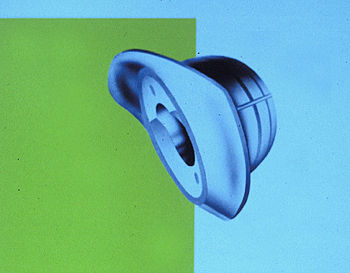 |
| Figure 3 : Elite | Figure 4 : Ogee Cup |
The stem-cement complex
Cement and heavy stems
"Heavy" and "extra heavy" stems (SR2 and SR3) were made by Thackray at the request of A.C.O.R.A. members, who wanted to have better fill of very wide femoral canals. In spite (or because) of advanced cementing, these stems failed rapidly, with proximal femoral loosening after 3 to 7 years. The components were rapidly abandoned. At the Lyons symposium, F. Michel summarized the Group’s experience as follows, "... in a large femoral cavity, it’s better to have cement than metal."
Cement and "standard" Charnley stems
Having a cement mantle that is thick and as even as possible is nowadays considered an absolute must, in the United States. At the first session of the Hip Society meeting in New Orleans, in March 1998,25 the Americans argued cogently in favour of pressurization, centralization, etc. (Padgett et al, Bartel et al, Maloney et al, Noble). Ling was the only one to suggest that making the cement completely non-porous could have detrimental effects - something that Hernigou had already suggested at the Lyons symposium. More recently, Joshi et al26 have shown clinical data demonstrating the link between the quality of cementing and the occurrence of osteolysis around original and Evolution stems implanted by Eftekhar (New York), between 1970 and 1985. Overall, osteolysis was found to be three times more common in men than in women; however, the incidence was three times less when the thickness of the cement mantle was 3 mm in all zones of the femur. This is why these authors recommend that the stem:canal ratio should be 60-70%, while the implant (= cement plus stem):canal ratio should be at least 99%.
With Charnley-Kerboull stems
These arguments run counter to the concept underlying the Charnley-Kerboull
(CMK) stems, which provide greater canal fill and, hence, a thinner cement
mantle. However, a study presented to SOFCOT by Sedel et al27 did not show any
significant differences in the mainly distal remodelling of bone in contact
with original Charnley stems and with Charnley-Kerboull stems: both patterns
were found to be well fixed after a follow-up of at least 10 years. This may
seem surprising, given the 50% greater bending stiffness of the bulkier CMK as
compared with the Charnley, and the fact that the Charnley probably also has a
thicker cement mantle. It would appear that - at least over the first 10
years, and in the two models of stainless steel stems discussed here - the
stem-cement complex behaves in the same way, regardless of the ratio between
the two components of the complex.
Thus, however forcefully the advocates
of maximum cementing may argue, the jury is still out on the question of how
much cement is actually required.
Femoral cementing techniques
Conventional (so-called first-generation) technique
The French call it "à la papa" (nice and leisurely), in English it is known as hand-packing: this is how the pioneers cemented the original Charnley flat-backs. With this technique, a maximum of cancellous bone was preserved. At the Lyons symposium, Picault acknowledged that, in retrospect, this empirical way of doing things had merit. Draenert presented more scientific evidence in favour of preserving bone in the medullary canal. First-generation cementing was used for the first 333 LFAs in the Mayo Clinic study. Very recently, the Mayo Clinic group published a study correlating the long-term outcome of the stems with the quality of initial cementing, judged on the appearance of debonding at the cement-implant interface in Gruen zone 1 during the first 5 years after the operation.28 A radiolucent line in this zone, indicative of early subsidence, was seen in one third of the 297 hips in the study. Stem survival using mechanical failure (revision of the femoral component because of aseptic loosening, radiographic loosening manifested by a fracture of the cement mantle, or a complete radiolucent line at the bone-cement interface) as the end point, was found to be statistically correlated with initial debonding. Implants that had no evidence of debonding on the 1-5-year radiographs had a probability of survival at 15 years of 91%; if the Zone 1 radiolucent line was > 2 mm, the probability was down to 50%. Debonding was not associated with pain in the hip. The authors warned that their results were obtained with a smooth, tapered, trapezoidal, collarless stem, and should not be extrapolated to cemented prostheses of different designs.
Advanced (second-generation) cementing
Madey et al29 felt that the use of a distal (plastic or bone) plug, and the introduction of the cement in a retrograde manner had markedly improved the outcome of the original Charnley stems in the Iowa University series. Of the 116 hips evaluated clinically and radiographically at 15 years, only 5% had femoral component loosening. However, in the 1998 annual report of the Swedish Hip Arthroplasty Register,14 use of this cementing technique plus pulsatile lavage of the femoral canal was found to have brought about only a slight improvement in the survival of Charnley stems implanted for OA. In the authors’ view, this failure to achieve more substantial improvement was due to the frequent use of outdated instruments up to 1994, and the virtually exclusive use in Sweden of a posterior approach, which is thought to carry a higher risk of component malpositioning. In the study by Kobayashi et al,13 newer cementing techniques did not bring about improved implant survival in patients with large width of the femoral canal.
Contemporary (third- and subsequent-generation) cementing
As far as can be ascertained, the use in LFA procedures of cement centrifugation, of centralization by means of a distal spacer (available with the Elite Plus), of proximal precoating, and of canal venting with transfemoral vacuum application (Draenert) has not, to date, been reflected in published clinical results.
Which cement?
While nothing is as yet known about the effects of contemporary cementing, the way in which Charnley THR survival is affected by the type of cement used has been studied. In the Norwegian Arthroplasty Register,30 high-viscosity cement with an antibiotic was found to give the best results, with a cumulative survival at 5.5 years of 98.7% (97.7% in the case of high-viscosity cement without antibiotics). Boneloc, however, was found to give the poorest results. Furnes et al31 found Boneloc cement to give inferior results both for Charnley prostheses and for Exeter prostheses. While there was a significant difference in the survival rates of the femoral components of the two devices (with the Charnley having an eight times higher revision risk), there was no significant difference between Boneloc-cemented Charnley and Exeter cups. Here, too, it would be dangerous to extrapolate from Charnley stems to cemented femoral components of different designs.
DESIGN EVOLUTION AND
CEMENTING
Charnley acetabular components
The cementless Charnley press-fit cup
Charnley used 336 cups of this design in the period from 1962 to 1065. Of the 169 cups discussed in Charnley’s first paper,1 6.5% were found to have tilted after 4 years, while only 1% of 210 cemented PE cups were loose. The press-fit version was, therefore, abandoned.
Survival of the standard cemented cup
The Wrightington cups reported on at the Lyons symposium had been studied
in a series published by Hodgkinson et al in 1993.32 In this study, the three
acetabular zones of DeLee and Charnley, and the senior author’s
classification, were used. At 10 years, out of 152 standard LFA cups, only 30%
had no demarcation, while 17% had definite loosening (Type 3 or 4). At Lyons,
Eftekhar presented the results he had published with Kobayashi and others:33
When studied with 10 to 20 years’ follow-up, 17% of the 328 original Charnley
cups had complete (Type 3) demarcation or migration (Type 4). Removal of
eburnated bone at the acetabular roof, and large acetabular angles, were risk
factors for demarcation in the OA group, while young patient age was found to
be a risk factor in the rheumatoid group. In a 1996 paper written with other
authors,34 Clarac, who had been at the Lyons symposium, gave the results of
309 cups implanted by himself. Using the presence of a radiolucent line as the
end point, acetabular component survival went from 81.5% at 10 years to 58% at
20 years. Using migration as the end point, survival went from 92.6% at 10
years to 75.2% at 20 years. Garcia-Cimbrelo et al had given a paper at Lyons;
these authors subsequently published their results, in 1997.35 They found a
cumulative probability of an original LFA cup not showing migration
(Hodgkinson Type 4 demarcation) at 20 years of 79%, while the cumulative
probability of a cup not being radiographically loose (Types 3 and 4
demarcation) was only 67%. As in the Mayo Clinic’s femoral component study,28
there was a strong correlation between the radiographic pattern during the
first years after the operation, and the survival of the cups. Thus, at the
time of the latest follow-up, migration was found in 5% of the cups with no
demarcation on the initial postoperative radiographs, while 35% of those with
Type 2 demarcation (Zones 1 and 2 lucency), and 72% of those with Type 3
demarcation (complete radiolucent line) had migrated. Kobayashi et al13
studied 293 LFAs (a mix of standard and Ogee cups), with an average follow-up
of 16 years; they found a rate of cup survival was 92.3% using revision as the
end point, and 83.6% when radiographic evidence of failure (Type 3 and 4
demarcation) was used as the end point.
The overall pattern that emerges
regarding the original cup is very uniform: at the same follow-up of 15 to 20
years, the cups do twice to three times less well than the femoral components
with which they articulate; and cup loosening is invariably correlated with
the quality of the acetabular bone stock.
The long posterior wall cup (1972)
Our literature search failed to produce any articles dealing specifically with this modification, which was introduced in 1972 and has remained a feature of the Charnley Evolution range ever since.
The offset-bore cup (1975)
This cup pattern was introduced by Charnley in 1975. The PE socket with an eccentrically placed cavity for the femoral head was designed to remedy the problem of insufficient PE thickness in the weight-bearing part of small (40- or 43-mm) sockets and in patients with deficient bone stock.36 At Wrightington, this cup was used in only 1.6% of the cases. Ioannidis et al37 reported the long-term radiological results of 58 THA using the offset-bore socket in dysplastic hips and in a few revision cases. At a mean follow-up of 9.8 years, the revision rate for aseptic migration was 5.2%.
The flanged Ogee configuration (1977)
With the introduction of the cement pressurization flange on the Evolution stem, Charnley, in 1977, launched the concept of the flanged PE cup, which led to the Ogee pattern, in 1982 (Fig. 4). The flanged rim was designed to pressurize the cement by restricting its extrusion from the acetabulum during cup insertion. The flange may be trimmed to size by the surgeon. The Wrightington results published in 199332 showed a 7.3% rate of loosening (Types 3 and 4) of the Ogee cup at 10 years; this rate was more than twice lower than that of the standard socket followed up for the same length of time. However, in the Wrightington series of 260 Ogee cups published by Sochart and Hardinge in 1998,20 cup survival using revision for aseptic loosening as the end point was 99% at 10 years, but fell to 84% at 15 years. In an attempt to improve the results, Mulier et al38 developed a high-pressure ("hydraulic") Ogee cup cementing technique, in 1984: The cup is fixed with screws through the flange, and the cement is injected through a hole, into the walled-off cavity between the cup and the bone. This allows the cement to polymerize behind a completely immobilized cup.
‘LFA-STYLE’
SOCKETS
The Wrightington cups: flanged and long posterior wall
Having abandoned trochanteric osteotomy and replaced the Charnley Evolution stem by the Wrightington FC device, Hardinge felt that a modification of the cemented cup was also indicated. In 1981, he designed his own flanged socket; the results obtained appear to be superior to those of the Ogee cup. A comparative study performed in 1981-8520 showed cup survival using revision for aseptic loosening as the end point of 99% at 10 years; by 15 years, this survival rate was still 95% (as against 84% for the Ogee cup). The 260 cups used in this study were not, however, of exactly the same design: 193 were all-PE Hardinge sockets, while 67 had a thin-section metal backing. This latter pattern had been introduced in 1985, no doubt under the influence of the studies by Harris et al, in order to improve the acetabular stress pattern and to prevent PE deformation. By 1997, none of these 67 cups had required revision.
The Charnley-Kerboull cup
It would appear that French orthopods using the LFA were not overly impressed with the acetabular components developed by Charnley across the Channel. The flanged socket design did not become popular in France. To the best of our knowledge, the socket of the CMK total hip prosthesis does not differ in any major way from the standard pattern of the original LFA. In the study by Dejean et al,22 there was a 45.7% rate of radiolucent lines at the cup/bone interface; in one third of the cases, the lucencies were progressive, leading, eventually, to 5 revisions (2 for loosening, 3 for wear), with an acetabular component revision rate of 7%. (With one fractured stem that was not revised, and 6 revised hips, the overall mechanical failure rate at 15-16 years in this series was thus 10%).
The Acoplot socket
This socket was launched in 1992, by the A.C.O.R.A. Group. It has convex pods, which, together with overreaming of the acetabulum to a diameter 2 mm greater than that of the definitive cup, provide for a cement thickness of 5 mm. At Lyons, Caton presented the first results, showing no revision, at 5 years, of the 58 Acoplot sockets in the study. In their very much earlier paper, Oh et al39 had investigated a number of cup surface designs, and recognized the utility of pods. Hodgkinson et al32 felt that such surface features would adversely affect cement pressurization and increase the production of PE wear debris at the socket/bone interface. This is why they opted for the Ogee pattern. Whatever the outcome of the argument over studs may be, recent work by Joshi et al26 suggests that an even cement thickness of 6 mm should be provided.
Cementless 22.2-mm sockets
Impressed by the hybrid THR concept advocated by Harris since the mid-80s,
and no doubt disappointed by the poorer survival of the cemented LFA cups as
compared with the stems, several LFA users experimented with hybrid versions
comprising a Charnley or Charnley-style stem and a variety of cementless
sockets. At Lyons, Prudhon presented a number of combinations (Saint-Nabor,
Aura, Alto, B2C), especially with Harris-Galante cups (a combination also used
by Önsten et al40 and by Callaghan in young patients). The Duraloc socket
features in the DePuy hybrid version of the Elite. In France, the A.C.O.R.A.
Group has, since 1995, been trying out a modular grit-blasted titanium cup
with a plasma-sprayed titanium coating.
The overwhelming majority of
manufacturers now have modular cementless sockets for their 22.2-mm
prostheses.
AND PARTICULATE DEBRIS
The 22.2-mm LFA in stainless steel-on-PE
At the Lyons symposium, there was agreement that PE wear was the most detrimental factor affecting the longevity of the Charnley LFA. Wear is mainly measured radiographically (Charnley, Livermore, etc.); none of the techniques devised to date are entirely accurate and reproducible.
Standard, LPW, and Ogee sockets
The mean annual PE wear rate of the original 22.2-mm stainless steel-on-PE
LFAs in the first series at Wrightington was 0.07 mm to 0.21 mm.36 In France,
a mean annual rate of 0.12 mm was found in the Cochin Hospital LFA study;4
however, in 50% of the cases in Clarac’s LFA study,34 the rate of wear was
higher. Using radiostereometry, a more accurate measuring technique, Önsten et
al40 found a mean annual wear rate of 0.09 mm at a mean follow-up of 5 years.
The same figure was reported by Dejean et al22 in a study of 70
Charnley-Kerboull THA at a mean follow-up of 15.6 years. (At the time of the
study, the Charnley-Kerboull was identical to the original LFA.) It follows
that a mean annual PE wear rate of 0.1 mm may be taken as the benchmark for
the 22.2-mm monobloc device with a stainless steel-on-PE combination.
The
wear rate may, however, by higher. In their tribological studies of original
LFAs retrieved because of mechanical problems (dislocation, loosening), Isaac
et al41 found that 25% of the cups had overall wear that could be as much as
3.5 mm after 2 to 7 years. They thought that hard particles of
roentgenographic contrast within the acrylic cement had scratched the polished
stainless steel heads. In extreme cases, progressive PE wear-out may lead to
cup fracture, as seen by Wroblewski et al43 in 7 LFAs that needed revision;
mean wear was found to have been 0.6 mm/year.
Kobayashi et al13, 33 are
convinced of the relationship between PE wear and implant loosening, with the
Charnley LFA. These authors identified the following risk factors for
excessive wear and, hence, loosening: young patient age, male gender, the type
of OA (normotrophic or atrophic), and socket cementing (cement mantle
deficiency in Zones I and II).
The relationship between PE wear and
osteolysis is equally obvious. Thus, Joshi et al26 reported a 15% osteolysis
rate in a total of 249 Charnley LFAs studied with more than 10 years’
follow-up. Where the criteria for cement mantle thickness had been met (6 mm
around the socket; 3 mm in all zones of the femur), the osteolysis rate was
only 5.2%. These results agree with those found by Garcia-Cimbrelo et al44 in
a study of femoral osteolysis in 680 LFAs at a mean follow-up of 16 years. In
the last-mentioned two papers, survivorship analysis showed a probability of
osteolysis of 1% (femur only) and 3% (femur and acetabulum) at 5 years; 6% at
10 years; 10% (f) and 11.3% (f + a) at 15 years; and 11% (f) and 17% (f + a)
at 20 years. This osteolysis, which in Garcia-Cimbrelo’s study occurred after
a mean of 9,3 years from arthroplasty, is always progressive.
Offset-bore sockets
In the Wrightington study reported on by Izquierdo-Avino et al,36 the mean annual wear rate of the 54 sockets studied was 0.04 mm, at a mean follow-up of 8.1 years. Rates approaching 0.06 mm in Zone I, and 0.04 mm in Zone II, were calculated in the series of 58 cups studied by Ioannidis et al.37 These rates are lower by a factor of 2 compared with those of the standard LFA sockets described above.
22.2-mm heads on Morse tapers
One of the last developments applied to the Elite Plus stem by Thackray was the introduction of a modular femoral component, with a 10-mm Morse taper. Since the take-over by DePuy in 1991, other head sizes (26, 28, and 32 mm) have also been available, as with all Charnley-style modular stems. Since PE wear is about twice less with ceramic heads than with metal (other than titanium) heads of the same size, ceramic-on-PE combinations have been introduced in the 22.2-mm size.
Alumina ceramic-on-PE
Many had thought that a 22.2-mm alumina ceramic head would be an impossibility; however, in 1988 Wroblewski et al45 started using a head made of that material to articulate with a cross-linked PE (XLP) cup. The top of the neck was parallel rather than tapered, with a 0.2-mm thick UHMPE sleeve between the neck and the head. After an initial bedding-in phase, with a mean penetration of 0.3 mm/year (18 months, 1.5 million cycles), wear decreased markedly and evend out at around 0.022 mm/year, which was 3 to 4 times less than the rate seen with the classical stainless steel-on-PE combination.
Zirconia ceramic-on-PE
This material is more widely used for femoral heads, and popular because of
its hardness. 22.2-mm diameter zirconia ceramic heads are available with the
Elite Plus, the A.C.O.R.A., and the Modulor femoral components. We have not
found any publications concerning the clinical results and in-vivo wear
measurements that could be used for a comparison with the 22.2-mm stainless
steel-on-PE combination.
The 22.2-mm titanium-on-PE combination used in the
CMKIII and the Capital prosthesis will not be discussed in this article, since
no results have been published on the former, and the sad tale of the latter
has already been recounted.
Wear debris and its toxicity
Metal debris
The production of metal debris is not confined to THA with metal bearing
surfaces. This kind of debris is seen mainly, though not exclusively, with
loose or worn implants. Case et al46 have shown chromosomal aberrations in
bone marrow cells adjacent to loose prostheses; of the implants examined, 27
(of 69) were Charnley THA. In 1997, the same team47 reported 4 new cases of
soft-tissue tumours in the vicinity of THAs, including one high-grade
leiomyosarcoma near a Charnley LFA to which the patient had been
revised.
Stainless steel and cobalt-chrome particles may also be spread
further afield by the bloodstream, and accumulate in lymphoreticular tissues
distant from the hip. Thus, metallic debris was found, at autopsy, in lymph
nodes, the liver, and the spleen of a patient who had received a Charnley
THA,48 and has also been reported in the bone marrow. 49 Little is known about
the long-term effects of this debris; however, this is a matter of some
concern, especially in younger patients.49
PE debris
It is now well known that particulate PE debris can cause local and regional damage, by triggering a cascade of cytochemical events leading to phagocytosis and osteolysis. However, PE debris has also been demonstrated in lymph nodes,50 especially in the pelvic nodes on the same side as the metal-on-PE THA.51 Nothing is known as yet about the eventual effects of these deposits; and further studies are required. There is, obviously, an urgent need for a PE material with greater wear resistance.
This article is intended to give an overview of the developments and modifications, over the past 35 years, of the Charnley low-friction arthroplasty. This survey should allow us to establish the "gold standard LFA", based upon implants with at least 20 years’ follow-up. It also shows the results in the general population. Anything done since 1986 has not yet been subjected to sufficiently long follow-up, and will require a few more years and, above all, published results, for its merits and demerits to be assessed. Any change made to the original concept can have unforeseeable repercussions, and the long-term results obtained with the original LFA cannot be extrapolated to other devices that are - more or less justifiably - described as "Charnley-style" THA, although, at times, the link with the original is no more than ... 7/8".
(Transl KRMB)
| REFERENCES Peer-reviewed international journals - 1993/1998 - Revues Internationales à Comité de Lecture : Rev Chir Orthop, J Bone Joint Surg (Am & Br), Clin Orthop, Acta Orthop Scand, J Arthroplasty, Orthopaedics, Intern Orthop (SICOT), Hip Intern (EHS) - French conferences: SOFCOT 1995, 1996, 1997 (Paris); Lyons International Symposium (December 1995) - International conferences: 65th Annual Meeting of the AAOS & the Hip Society 1998 (New Orleans); European Hip Society 1996 (Helsinki) and 1998 (Beaune) 1. Charnley J : The long-term results of low-friction arthroplasty of the hip performed as a primary intervention. J Bone Joint Surg 54B: 61, 1972. Reedited in Clin Orthop 319: 4, 1995 2. Isaac GH, Wroblewski BM, Atkinson JR, Dowson D : Source of the cement within the Charnley hip. J Bone Joint Surg 72B: 149, 1990 3. Charnley total hip arthroplasty. 33 years of world-wide experience. Directed by J. Caton, F. Michel, C. Picault. Edited by the A.C.O.R.A. Group. TRANSIT communications. N.H.A.-Lyon, 1995 4. Wicart P, Dejean O, Kerboull M : Prothèses totales de hanche de Charnley : étude clinique et radiographique de 15 à 25 ans après leur implantation. Rev Chir Orthop 82(suppl II): 76, 1996 5. Marduel-Picault MS : Devenir à plus de 20 ans des prothèses totales de hanche de type Charnley. Thèse N°254, Université Claude Bernard Lyon I, 1996 6. Ranawat C, Hansraj K, Msika C, Neves M : Etude de survie d’une série de prothèses totales de hanche de type Charnley. Rev Chir Orthop 79(suppl I): 130, 1993 7. Karachalios T, Hartofilakidis G, Zacharakis N, Tsekoura M : A 12- to 18-year radiographic follow-up study of Charnley low-friction arthroplasty. The role of the center of rotation. Clin Orthop 296: 140, 1993 8. Kavanagh BF, Wallrichs S, Dewitz M, Berry D, Currier B, Ilstrup D, Coventry MB : Charnley low-friction of the hip. Twenty-year results with cement. J Arthroplasty 9: 229, 1994 9. Neumann L, Freund KG, Harry Sorenson K : Long-term results of Charnley total hip replacement. Review of 92 patients at 15 to 20 years. J Bone Joint Surg 76B: 245, 1994 10. Garellick G, Herberts P, Strömberg C, Malchau H : Long-term results of Charnley arthroplasty. A 12-16-year follow-up study. J Arthroplasty 9: 333, 1994 11. GARDES P., FAVARD L., GARDES JC : Révision à long terme d’une série homogène et consécutive de 100 prothèses totales de hanche type “Charnley”. Rev Chir Orthop 82: 306, 1996 12. Hozack WJ, Rothman RH, Booth RE, Rao R, Eng K : Twenty year survivorship of 1,157 Charnley cemented total hip arthroplasties. J Arthroplasty 12: 219, 1997 13. Kobayashi S, Takaoka K, Saito N, Hisa K : Factors affecting aseptic failure of fixation after primary Charnley total hip arthroplasty. J Bone Joint Surg 79A: 1618, 1997 14. Malchau H, Herberts P : Prognosis of total hip replacement. Revision and re-revision rate in THR : a revision-risk study of 148,359 primary operations. Scientific exhibition, 65th annual meeting of the AAOS, New-Orleans, February, 1998 15. Soyer J, Avedikian J, Pries P., Clarac JP : Comportement à long terme de l’implant fémoral de Charnley. Revue de 309 dossiers avec un recul minimum de 20 ans. Rev Chir Orthop 83: 416, 1997 16. Garcia-Cimbrelo E, Madero R, Blasco-Alberdi A, Munuera L : Femoral osteolysis after low-friction arthroplasty. A planimetric study and volumetric estimate. J Arthroplasty 12: 624, 1997 17. Raut VV, Siney PD, Wroblewski BM : Long-term results of cemented revision arthroplasty for fractured stem. Clin Orthop 303: 165, 1994 18. Wroblewski BM : Charnley low friction arthroplasty. Review of the past, present status and prospects for the future. Clin Orthop 210: 37, 1986 19. Dall DM, Learmonth ID, Solomon MI, Miles AW, Davenport JM : Fracture and loosening of Charnley femoral stems. Comparison between first-generation and subsequent designs. J Bone Joint Surg 75B: 259, 1993 20. Sochart DH, Hardinge K : Comparison of the Wrightington FC hip with the Charnley low-friction arthroplasty. 10- to 15-year results and survival analysis. J Bone Joint Surg 80B: 577, 1998 21. Kerboull M : La prothèse Charnley-Kerboul. In Arthroplastie totale de hanche. Ed: M. Postel, M. Kerboul, J. Evrard, JP. Courpied. Springer-Verlag Ed., Berlin, Heidelberg, New-York, Tokyo; p.14 - 19, 1985 22. Dejean O, Kerboull L, Kerboull M : Devenir à plus de 15 ans des prothèses totales de hanche de type Charnley. Rev Chir Orthop 81(suppl I): 142, 1995 23. Langlais F : Tolérance des prothèses totales de hanche en alliage de titane. Ann Orthop Ouest 25: 61, 1993. 24. Massoud SN, Hunter JB, Holdsworth BJ, Wallace WA, Julusson R : Early femoral loosening in one design of cemented hip replacement. J Bone Joint Surg 79B: 603, 1997 25. Cemented femoral fixation - issues for the twilight of the twentieth century: Proceedings of The Hip Society: p.14 -21, New Orleans, 1998 26. Joshi RP, Eftekhar NS, McMahon DJ, Nercessian OA : Osteolysis after Charnley primary low-friction arthroplasty. A comparison of two matched paired groups. J Bone Joint Surg 80B: 585, 1998 27. Sedel L, Meunier A, Gaucher F, Pasquier G, Souchet M, Kerboull M, Witvoet J : Remaniements osseux à long terme au contact de prothèses fémorales cimentées. Rev Chir Orthop 79(suppl I): 129, 1993 28. Berry DJ, Harmsen WS, Ilstrup DM : The natural history of debonding of the femoral component from the cement and its effect on long-term survival of Charnley total hip replacements. J Bone Joint Surg 80A: 715, 1998 29. Madey SM, Callaghan JJ, Olejniczak JP, Goetz DD, Johnston RC : Charnley total hip arthroplasty with use of improved techniques of cementing. The results after a minimum of fifteen years of follow-up. J Bone Joint Surg 79A: 53, 1997 30. Havelin LI, Espehaug B, Vollset SE, Engesaeter LB : The effect of the type of cement on early revision of Charnley total hip prostheses. A review of eight thousand and seventy nine primary arthroplasties from the Norvegian Arthroplasty Register. J Bone Joint Surg 77A: 1543, 1995 31. Furnes O, Lie SA, Havelin LI, Vollset SE, Engesaeter LB : Exeter and Charnley arthroplasties with Boneloc or high viscosity cement. Comparison of 1.127 arthroplasties followed for 5 years in the Norvegian Arthroplasty Register. Acta Orthop Scand 68: 51, 1997 32. Hodgkinson JP, Maskell AP, Paul A, Wroblewski BM : Flanged acetabular components in cemented Charnley hip arthroplasty. J Bone Joint Surg 75B: 464, 1993 33. Kobayashi S, Eftekhar NS, Terayama K, Ioro R : Risk factors affecting radiological failure of the socket in primary Charnley low friction arthroplasty. A 10- to 20-year followup study. Clin Orthop 306: 84, 1994 34. Avedikian J, Soyer J, Dumez JF, Muller A, Pries P, Clarac JP : L’implant cotyloïdien dans l’arthroplastie totale de Charnley. Revue de 309 dossiers avec un recul minimum de 15 ans. Rev Chir Orthop 82: 116, 1996 35. Garcia-Cimbrelo E, Diez-Vazquez V, Madero R, Munuera L : Progression of radiolucent lines adjacent to the acetabular component and factors influencing migration after Charnley low-friction total hip arthroplasty. J Bone Joint Surg 79A: 1373, 1997 36. Izquierdo-Avino RJ, Siney PD, Wroblewski BM : Polyethylene wear in the Charnley offset bore acetabular cup. A radiological analysis. J Bone Joint Surg 78B: 82, 1996 37. Ioannidis TT, Zacharakis N, Magnissalis EA, Eliades G, Hartofilakidis G : Long-term behaviour of the Charnley offset-bore acetabular cup. J Bone Joint Surg 80B: 48, 1998 38. Mulier M, Van Oost J, Van Lauwe J, Feron M, Bellon E, Suetens P : Modified technique of cementing the Charnley cup. Scientific Exhibit 064, 68th Congress of the AAOS, New-Orleans, March 1998 39. Oh I, Sander TW, Treharne RW : Acetabular cup groove and pod design and its effect on cement fixation in total hip arthroplasty. Clin Orthop 189: 308, 1984 40. Önsten I, Carlsson AS, Besjakov J : Wear in uncemented porous and cemented polyethylene sockets. A randomised, radiostereometric study. J Bone Joint Surg 80B: 345, 1998 41. Isaac GH, Wroblewski BM, Atkinson JR, Dowson D. : A tribological study of retrieved hip prostheses. Clin Orthop 276: 115, 1992 42. Wroblewski BM, Siney PD : Charnley low-friction arthroplasty of the hip. Long-term results. Clin Orthop 292: 191, 1993 43. Wroblewski BM, Siney PD, Fleming PA : Wear and fracture of the acetabular cup in Charnley low-friction arthroplasty. J Arthroplasty 13: 132, 1998 44. Wroblewski BM, Siney PD, Dowson D, Collins SN : Prospective clinical and joint simulator studies of a new total hip arthroplasty using alumina ceramic heads and cross-linked polyethylene cups. J Bone Joint Surg 78B: 280, 1996 45. Case CP, Langkamer VG, Howell RT, Webb J, Standen G, Palmer MR, Kemp AJ, Learmonth ID : Preliminary observations on possible premalignant changes in bone marrow adjacent to worn total hip arthroplasty implants. Clin Orthop 329S: S269, 1996 46. Langkamer VG, Case CP, Collins C, Watt I, Dixon J, Kemp AJ, Atkins RM : Tumors around implants. J Arthroplasty 12: 812, 1997 47. Langkamer VG, Case CP, Heap PF, Taylor A, Collins C, Pearse M, Solomon L : Systemic distribution of wear debris after hip replacement. A cause of concern ? J Bone Joint Surg 74B, 831, 1992 48. Case CP, Langkamer VG, James C, Palmer MR, Kemp AJ, Heap PF, Solomon L : Widespread dissemination of metal debris from implants. J Bone Joint Surg 76B: 701, 1994 49. Shea KG, Bloebaum RD, Avent JM, Birk GT, Samuelson KM : Analysis of lymph nodes for polyethylene particles in patients who have had a primary joint replacement. J Bone Joint Surg 78A, 497, 1996 50. Hicks DG, Judkins AR, Sickel JZ, Rosier RN, Puzas JE, O’Keefe RJ : Granular hystiocytosis of pelvic lymph nodes following total hip arthroplasty. J Bone Joint Surg 78A, 482, 1996 |